Needle holder grasps in surgery

For the spay-neuter vet, pandemic social distancing has been a slow time. I’ve had plenty of opportunity to stay home sleeping, baking, playing Pokemon Go and watching birds (often these two are simultaneous activities), eating fiddleheads, and seeing spring unfurl. With services starting to reopen, I’m getting ready to go back to work on Monday, so my thoughts have started to turn back to surgery and ergonomics.

During these idle months I’ve had some time to look through old ergonomics articles and projects with an eye to assembling them into something useful. This week I found myself thinking about needle holder grasps (after a Facebook conversation) and thought to look back at my masters thesis in ergonomics. The topic was a comparison between palm grasp and tripod (fingers in the rings) grasp for needle holders. The aim was to compare users of the two techniques both by surveying them about pain and by measuring the muscular strain in their forearms. In keeping with my desire to share incidental and independent research results, I’m publishing the thesis at the bottom of this blog post (never fear, we were limited to 5000 words).

If you thought this article would answer the question of whether palm grasp is better than placing fingers in the rings of the instrument, think again! The utter messiness of the results and the difficulty of drawing conclusions about the different grasp types was why I never published it anywhere (until now! here!). But I learned a lot from the research about individual variability and the diversity of supposedly standardized techniques. This paragraph taken from the Discussion sums up what I learned:

The amount of grasp diversity between participants, the use of non-standard instrument grasps, and the variability in individual participants’ grasps, were surprising findings in the current study. Participants were often unaware of the grasp that they used. Several described themselves using a grasp different from the one that they actually used, and some noticed in the midst of the experiment that they were using a different grasp from what they had described. Even within a single grasp type, the participants varied in their movement patterns and degrees of forearm rotation and wrist angulation. 

So while I can’t promise any groundbreaking information about which grasp style is ergonomically superior, I do think there are some interesting photos and tidbits in this article. I also think that the diversity of successful techniques leaves clear opportunity for individuals to modify their grasping and suturing technique if and when it becomes painful or problematic for them.

I hope you all are well, and staying safe. Enjoy!

Electromyographic analysis of needle-holder grasps used while suturing

A thesis in partial fulfillment of Masters of Science in Health Ergonomics, University of Derby, February, 2015

Abstract

The current study examined variations in muscular force and muscle use patterns between surgeons using different grasps while suturing. Fourteen (4 male, 10 female) right-handed veterinarians were recruited into one of two groups, palm or tripod, depending on their usual, preferred needle holder grasp. Participants completed the Cornell Musculoskeletal Discomfort Questionnaire (CMDQ) and the Cornell Hand Discomfort Questionnaire (CHDQ), and then performed a suturing task using their preferred grasp. Four muscles in the right forearm region were selected for electromyographic (EMG) recording: extensor digitorum communis (EDC), flexor carpi radialis (FCR), flexor carpi ulnaris (FCU) and abductor pollicis longus/ extensor pollicis brevis (APL). 

The 1-week period prevalence of MSD was 92.9%, with 13 of 14 participants reporting pain. Of these, 7 (50%) reported hand pain, and 12 (85.7%) reported body pain. Observed grasps differed from those reported by participants, with five using exclusively tripod grasp with the thumb and ring finger in the instrument rings, two using a modified tripod grasp with thumb and middle finger, three using palm grasp for suture placement and tripod grasp during needle extraction and knot tying, three using palm grasp with no fingers in the instrument rings, and one using palm grasp with the ring finger in one instrument ring. The static load (10th% APDF) on each of the four muscles ranged from 0.9 to 10% MVC, with greater mean values for the extensor EDC than for the flexors FCR and FCU. Degrees of forearm pronation and supination ranged from 80 to 180 degrees, and degree of rotation was positively correlated with the total pain score. Future investigation into the characteristics and benefits of various grasps is warranted, so that practical advice on reducing strain and MSD risk can be offered to surgeons.

Introduction 

High-volume spay-neuter is a growing practice area in veterinary medicine in the US (Looney et al., 2008) in which veterinarians may perform over 30 surgical procedures daily, and some individuals spend over 35 hours each week performing surgery (White, 2013). These procedures are of limited variety compared with general surgery, and frequently involve static postures and repetitive manual tasks. Repetitive work is associated with increases in upper limb discomfort, tendinitis, and carpal tunnel syndrome in people who engage in manual work (Latko et al., 1999), and static postures, or isometric positions where little movement takes place, multiply the risk for musculoskeletal disorders attributable to those postures (Esser et al., 2007). While work in high volume spay-neuter has many qualities that would appear to put veterinarians at risk for MSD, there is limited research on the effects of this repetitive surgical workload on veterinarians, and no research exploring ways to mitigate these effects. 

A single cross-sectional study (White, 2013) has investigated musculoskeletal discomfort (MSD) in veterinarians working in high-volume spay-neuter. The one-month period prevalence of MSD was 99.1%, with 76.7% experiencing hand or wrist pain and 98.2% experiencing body pain. Hand discomfort was most commonly reported in the right thumb and/or thumb base (49.8%) and the right wrist (37.9%). This rate of hand/wrist discomfort is 1.5 to 2 times the prevalence of upper limb MSD experienced by veterinarians in general practice (Kozak et al., 2014; Scuffham et al., 2010; Smith et al., 2009), and greater than the prevalence in surgeons in human surgical practice (Adams et al., 2013; Soueid et al., 2010). Body discomfort in spay-neuter veterinarians was most commonly reported in the lower back (76.7%), shoulders (72.6%), and neck (71.7%). Three-quarters of veterinarians experiencing hand, finger, and thumb MSD attributed their MSD completely to their work in spay-neuter. Increasing career length, increasing weekly hours in surgery and decreasing job satisfaction were the work-related factors with the greatest relative contribution accounting for variation in hand pain severity and total pain. While 94.4% of respondents felt that posture and positioning during surgery is important, only 30.6% had received any instruction in posture, positioning, or ergonomics in surgery (White, 2013).

The high prevalence of upper limb MSD in spay-neuter veterinarians may be related to the high volume and limited variety of surgical tasks undertaken, and thus the repetition of a limited diversity of hand movement patterns performed in the workday. Anecdotally, some spay-neuter veterinarians have attributed their lack of upper limb MSD to their use of a palm grasp when using needle holders, instead placing their fingers in the instrument rings. 

Textbooks and authors vary in their use of terms to describe instrument grip. Anderson and Romfh (1980) describe the “palmed grip” in which the surgeon grips a long needle holder by the shanks, away from the finger rings and ratchet, making it impossible to open or close the ratchet while using this grip. This is in contrast to Seki’s (1988) diagram of “grip 2,” in which the finger rings and ratchet are held in the palm of the hand, allowing operation of the ratchet mechanism. This is the same as the “modified thenar eminence grip” described by Toombs and Bauer (1993), and also described (though unnamed), two decades earlier (Weiss, 1973). More recent sources (Kirpensteijn & Klein, 2006; Yoon & Mann, 2011) name this same grasp the “palm grip.” Yoon and Mann (2011) use the term “thenar eminence grip” to describe a grasp in which the needle holder is grasped in the palm, with the tip of the ring finger placed through one finger ring. The same grasp is elsewhere called the “thenar grip” (Anderson & Romfh, 1980). Booth (2013) repeats the descriptions and terms used by Anderson and Romfh (1980), except that, in the illustration of Booth’s “thenar grip,” the fourth finger does not enter the finger ring, making this “thenar grip” resemble the “palm grip” described above.

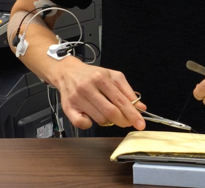
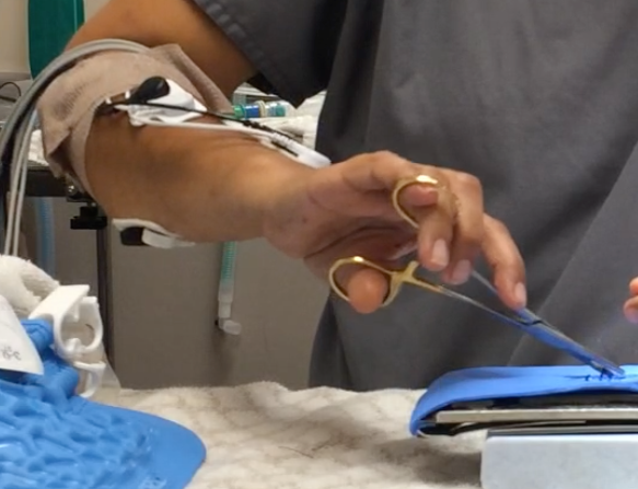
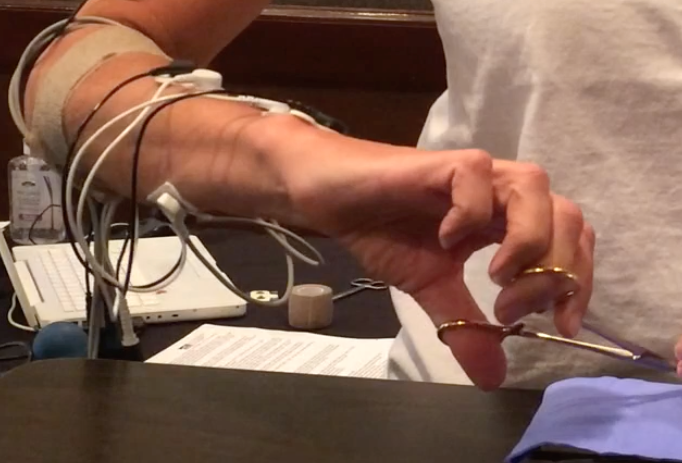
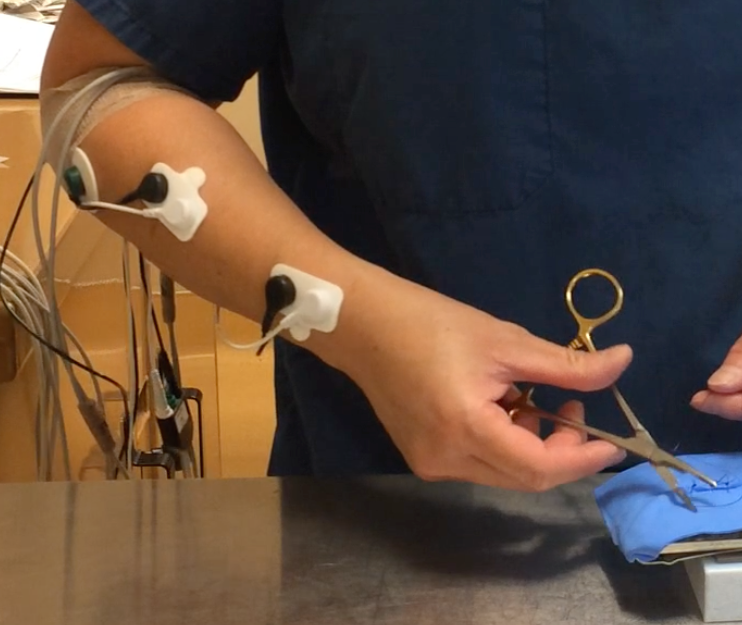
Current consensus appears to favor “palm grip” to describe the grasp in which the finger rings and ratchet are held in the palm, with no fingers in the finger rings. The comparison grip, utilizing thumb and ring finger in the instrument rings, has been called the three point grip (Kirpensteijn & Klein, 2006), the thumb-ring finger grip (Anderson & Romfh, 1980), the thumb-third finger grip (Toombs & Bauer, 1993) [this grip is pictured with the fourth phalanx in the instrument ring, despite the use of “third finger” in the name], and thumb-ring finger (tripod) grip (Booth, 2013). For the current study, the term “tripod grip” has been chosen for its brevity and clarity.

The research comparing the attributes, physics, and precision of these grasps during open (non-laparoscopic) surgery is limited. One study found greater suturing precision among surgeons using palm grasp as compared to tripod grasp (Seki, 1988), and the author speculated that the palm grasp was more stable and reduced the difference in angle between the hand and the instrument. Despite the limited research comparing the grasps, surgical textbooks make assertions about their qualities and disadvantages. Toombs and Bauer (1993) state that the modified thenar eminence (palm) grip results in imprecise release of the needle, making this grip poorly suited to delicate suturing compared to the thumb-third finger (tripod) grip. This contrasts with Seki’s (1988) finding of greater accuracy when using the palm grip.

Several studies have used electromyography (EMG) to compare the ergonomic aspects of various grasps. Surface EMG uses electrodes on the skin to detect the electrical activity produced by the summed motor unit action potentials in the muscle of interest (Criswell, 2011). EMG signal strength has an approximately linear relationship with muscular force, making it useful in ergonomics for determining the amount of individual muscle involvement in a given task, and allowing evaluation of strain on the tissues (Hägg et al., 2004). One study comparing two different grasp styles on a laparoscopic instrument found differences in EMG amplitude in several forearm muscle groups, leading to the recommendation of a specific, palm-grasp style in certain circumstances (Berguer et al., 1999). A second study comparing various laparoscopic handle designs found that the pattern of EMG activity—the proportional use of each measured muscle—is characteristic of the handle (and thus the grasp) used, rather than being task-specific (Matern et al., 2004). A similar EMG study of handwriting grasp styles also showed characteristic EMG activation patterns for each grasp style (de Almeida et al., 2013).

The amplitude probability distribution function (APDF) is a means of EMG data reduction that is used to characterize the muscular load profile over a period of time (Hägg et al., 2004). The calculation reveals the cumulative probability for EMG amplitude over time, and can be normalized for each subject to a percentage of their maximum voluntary contraction (%MVC) for that muscle. APDF levels are often reported as 10th, 50th, and 90th percentiles, with 10th %APDF representing static load, 50th% APDF median load, and 90th% APDF considered peak load for that muscle (Szeto et al., 2009). 

The current study aimed to examine variations in muscular force and muscle use patterns between surgeons using a palm grasp versus a tripod grasp while suturing. It was expected that the results could be used to guide surgeons in selecting which grasp to use routinely, and indicate which grasp to choose or avoid to decrease strain on specific muscles and their associated tendons and ligaments.

Methods

Participants 

A total of 14 (4 male, 10 female) veterinarians were recruited for the present study. Participants were recruited into one of two groups, palm or tripod, depending on their usual, preferred needle holder grasp pattern. All subjects were right-handed. 

Participants were recruited at two veterinary conferences: the North American Spay/Neuter Conference in Austin, Texas and the Silicon Valley Spay & Neuter Symposium in Milpitas, California, in 2014. Two additional veterinarians were recruited at a spay/neuter strategy meeting in Burlington, Vermont. Consent to participate was obtained from each participant before the study began. The study was approved by the Psychology Research Ethics Committee at the University of Derby

All participants in the study were asked to complete a questionnaire prior to participation. Demographic data including year of birth, year of graduation from veterinary school, whether they have obtained any specialty certification, and current hours per week performing surgery were recorded. Participants were shown pictures of “palm” and “tripod” grasps, and asked which grasp they use, or, if a mixture of grasps, in what proportion they use those grasps. They were also asked when they began using their current instrument grasp, whether they have used a different grasp at any point in their career, and why they have chosen their current instrument grasp.

In addition, participants were asked to complete the Cornell Musculoskeletal Discomfort Questionnaire and the Cornell Hand Discomfort Questionnaire regarding any discomfort in the past week. These questionnaires allowed the determination of the location, severity, and impact on work and daily activities.

Electromyography

Four muscles in the right forearm region were selected for the electro- myography (EMG) study: extensor digitorum communis (EDC), flexor carpi radialis (FCR), flexor carpi ulnaris (FCU) and abductor pollicis longus/ extensor pollicis brevis (APL). The I-330-C2+ system (J&J Engineering, Inc., Poulsboro, WA) was used to capture the surface EMG data, with a bandwidth of 10-400 Hz and a common mode rejection ratio of 100 dB, with input impedance 10 GW and a notch filter of 60 Hz. The EMG signals underwent a 16 bit analogue to digital (A/D) conversion at a sampling frequency of 1024 Hz.

Bipolar Ag-AgCl surface electrodes (Norotrode 20, Myotronics, Inc, Kent, WA) with an inter-electrode spacing of 22 mm were used. The ground electrode was an 1 3/8 inch Ag-AgCl electrode (SilveRest, Vermed, Bellows Falls, VT) that was placed on the right upper arm above the elbow.

The locations for EMG electrodes were adopted from Perotto (2011) and Criswell (2011). Before attaching electrodes, the skin was prepared by abrading with a gauze sponge. After electrode placement, the skin impedance was checked using the impedance testing function in the I-330-C2+, and impedance below 900 KW was considered acceptable. 

Prior to starting the experiment, subjects were asked to perform two trials of resisted isometric maximum voluntary contractions (MVC) of 5 seconds each against manual resistance for each muscle. 

Video Recording

Each session was recorded using 1080p HD video at 30 frames per second using an iPhone 5S (Apple, Inc, Cupertino, CA). A single, front view recording of each participant was made, and markers in the EMG recording allowed synchronization of video and EMG recordings. 

Video recordings were used to examine posture during surgery using Rapid Upper Limb Assessment  (McAtamney & Corlett, 1993). RULA assessment was made at the time in the work cycle when the highest loads occurred, assessing the participant’s dominant arm, and applied just to the experimental condition (not extrapolated to a “typical” work day).

Protocol

Each participant stood at a table adjusted to their preferred height. Participants were then asked to use an 5.5 inch Olsen-Hegar needle holder (Spectrum Surgical, Stow, OH) and thumb forceps to place five simple interrupted sutures in a polyvinyl alcohol synthetic chamois skin model using 3-0 Monocryl suture on a 40 mm, ½ circle taper needle. Measurements taken during the first suture were not included in the analysis, in order to allow the surgeon to become familiar with the materials and task.

Data Processing and Analysis

The USE3 Physiolab (J&J Engineering, Inc., Poulsboro, WA) software was used to process the EMG data. Data processing involved full-wave rectification and smoothing with root-mean-square (RMS) with a 250 ms window. These data were then exported to Microsoft Excel to compute the MVCs for each muscle, and to SPSS to compute the 10th%, 50th% and 90th% levels of Amplitude Probability Distribution Function (APDF) for the four muscle groups. 

            Pain severity for each body region was calculated for each participant using the scoring guidelines accompanying the CMDQ and CMHQ (Hedge, n.d.). Frequency scores were assigned: never = 0; 1–2 times a week = 1.5; 3-4 times a week = 3.5; every day = 5; several times a day = 10. Discomfort scores were assigned: slightly uncomfortable = 1; moderately uncomfortable = 2; very uncomfortable = 3. Daily interference scores were assigned: not at all = 1; slightly interfered = 2; substantially interfered = 3. Pain severity was obtained by multiplying the frequency, discomfort, and interference scores for each body part. Total body pain severity for an individual was obtained by summing all the body pain severity scores for that individual. Total hand pain scores were obtained by summing the hand pain severity scores for that individual. Total overall pain scores were obtained by summing the hand pain and body pain scores for that participant. 

            Demographic, MSD, and EMG data were entered into SPSS. Pearson correlations were used to assess relationships between MSD and demographic and postural characteristics. APDF levels of different muscles were compared using paired sample t-tests.

Results

Demographics

            A total of 14 veterinarians participated in this study, including 10 (71.4%) females and 4 (28.6%) males (Table 1). The median age of participants was 43 years, with a range of 31 to 62 years of age. Median time since graduation from veterinary school was 13.5 years, with a range of 4 to 32 years. None of the veterinarians had obtained board specialty certification. Participants spent a median of 17.5 hours a week in surgery, with a range from 0 to 35 hours weekly. Two participants did not regularly perform surgery in their current jobs: one was in a management position and performed surgery on an as-needed basis, and the other was seeking employment. Both of these veterinarians had several years experience performing surgery.

Table 1. Participant demographic, workload, instrument grip, and discomfort data

Musculoskeletal Discomfort Prevalence 

The self-reported 1-week period prevalence of MSD was 92.9%, with 13 of 14 participants reporting pain. Of these, 7 (50%) reported hand pain, and 12 (85.7%) reported body pain. All who reported discomfort also reported that it interfered at least slightly with their ability to work. In the right hand, the most commonly reported areas of MSD were the distal thumb (first proximal and distal phalangeal area; 28.6%), and the thumb base (first metacarpal area; 28.6%). MSD was reported in some portion of the right thumb [phalangeal and metacarpal areas] by 42.8% of participants. Body MSD was most commonly reported in the lower back (71.4%), right shoulder (50%), and neck (50%). 

Pain severity was not correlated with age (r (12)= 0.233, p= 0.424) or hours per week in surgery (r (12)= 0.005, p= 0.987), and was unrelated to the sex of the participant (t(12) = -1.415, p=0.182).

Grasp Characteristics

            Eight of the participants reported using tripod grasp all or most of the time, and 5 reported using palm grasp all or most of the time. The remaining surgeon reported using the two grasps equally. Eleven participants (78.6%) reported having adopted their current grasp in veterinary school or before, whereas 3 participants (21.4%) reported to have modified their grasp after graduation from school.

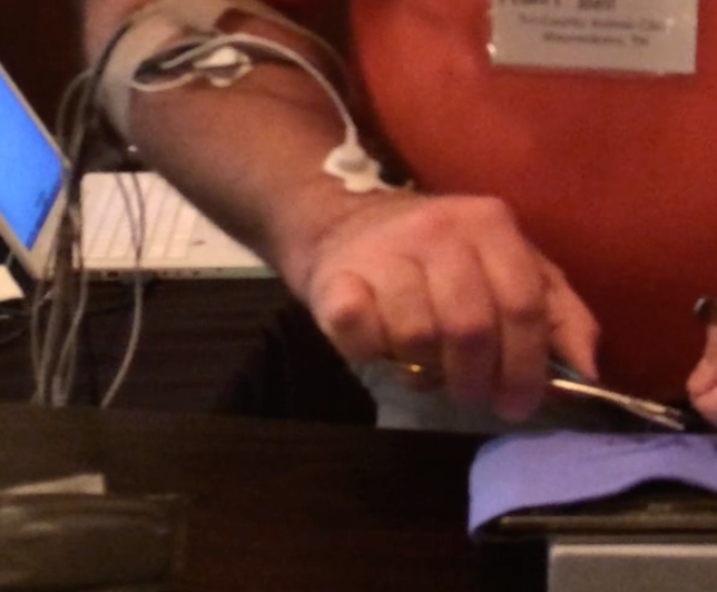
Actual observed grasps differed from those reported by participants (Figures 1-5). Video analysis revealed that 5 participants used exclusively tripod grasp with the thumb and ring finger in the instrument rings (1, 4 tripod), one of whom routinely placed her fifth finger in the instrument ring with her fourth finger. Two participants used a modified tripod grasp with the thumb and middle finger (1, 3 tripod) in the instrument rings. Three participants used palm grip for suture placement (driving the needle through the substrate) and switched to 1, 4 tripod grasp during needle extraction and knot tying (palm/tripod). Three participants used palm grasp with no fingers in the instrument rings, and one participant used a palm grasp with the ring finger in one instrument ring.

Figure 1. Instrument grasps used by study participants.: 1,4 tripod grasp
Figure 2. Instrument grasps used by study participants: 1,4 tripod grasp with fifth finger in ring.
Figure 3. Instrument grasps used by study participants: 1,3 tripod grasp
Figure 4. Instrument grasps used by study participants: Palm grasp.
Figure 5. Instrument grasps used by study participants: Palm grasp with fourth finger in ring.

Electromyography

Results of electromyographic recordings are presented in Table 2. In one participant, EMG readings were not obtained for FCU activity, as the electrodes loosened during the experiment.

Table 2. Results of low (10th % APDF), median (50th % APDF), and high (90th % APDF) muscle activities for all muscle groups and each participant. All values are expressed as a percentage of the maximum voluntary contraction (%MVC) for that muscle in that participant.

EDC: extensor digitorum communis, FCR: flexor carpi radialis, FCU: flexor carpi ulnaris, APL: abductor pollicis longus/ extensor pollicis brevis. 
*FCU electrodes loosened on Participant 7, preventing data collection from this muscle

The 10th % APDF, representing the static load on each of the four muscles, ranged from 0.9 to 10% MVC, with greater mean values for the extensor EDC (M = 5.51; SD = 1.37) than for the flexors FCR (M = 3.27, SD= 1.62) and FCU (M = 3.33, SD=1.53). These differences were statistically significant, with EDC:FCR t(13) = 5.082, p<0.001 and EDC:FCU t(12) = 4.824, p<0.001, two tailed. This differential activation level persisted between the EDC and FCU at the 50th and 90th % APDF, whereas the mean activity level of the FCR increased by the 50th and 90th % APDF so that there was no difference at either time between EDC and FCR activation levels (Figure 6). 

Figure 6. Muscle activation of extensors and flexors at the 10th, 50th, and 90th percentile APDF, expressed as a percentage of the maximum voluntary contraction (%MVC) for that muscle.

EDC: extensor digitorum communis, FCU: flexor carpi ulnaris, FCR: flexor carpi radialis. 
* significant difference between mean activation levels.

The unexpected diversity of grasp styles and small number of participants using each grasp prevented adequate comparisons of muscle activation patterns between grasps.

Postural comparisons

RULA assessments produced scores of 3 or 4 in all subjects, indicating that overall postural scores did not differ substantially between subjects, and that all fell into the moderate risk category. Variations in the degrees forearm pronation and supination were noted between subjects, with a range of 80 to 180 degrees of rotation (M= 125, SD = 26.5). The degree of rotation did not appear to be related to the instrument grasp, and was positively correlated with the total pain score determined on the CMDQ and CHDQ questionnaires (r (12)= 0.556, p= 0.039).

Discussion

There has been little previous research into the physical demands of high volume spay neuter surgery. A previous study of MSD prevalence in spay neuter veterinarians (White, 2013) found a 99.1% one-month period prevalence of MSD, which is slightly higher than the 92.9% one-week period prevalence reported in the current study. The body sites with the highest prevalence of MSD were the same in the two studies, with participants most often reporting body MSD in the lower back, shoulders and neck, and hand MSD in the right distal thumb and in the right thumb base. The previous study demonstrated increased MSD risk with increased weekly surgery hours and increased years of work, an effect not seen in the current study. However, these factors had weak explanatory power, accounting for less than 5% of the variability in MSD scores. This small effect size, paired with the smaller sample size in the current study, may account for this lack of effect. Neither study showed an effect of gender on MSD prevalence.

The EMG findings of greater static load on extensors compared to flexors is likely due to the extension of the metacarpophalangeal joints required to execute any of the needle holder grasps. During median and high load conditions, greater need for wrist flexion increases flexor load. Some of the increase with load in the FCR readings may also be due to crosstalk with the superficial digital flexor (Criswell, 2011), and may be related to creating a tighter instrument grasp as greater force is required.

The amount of grasp diversity between participants, the use of non-standard instrument grasps, and the variability in individual participants’ grasps, were surprising findings in the current study. Participants were often unaware of the grasp that they used. Several described themselves using a grasp different from the one that they actually used, and some noticed in the midst of the experiment that they were using a different grasp from what they had described. Even within a single grasp type, the participants varied in their movement patterns and degrees of forearm rotation and wrist angulation. 

Most of the participants claimed to have used their current instrument grasp beginning in veterinary school. However, the wide diversity of grasps, and the use of grasps not described in most veterinary or surgical texts, suggests either that the participants modified their grasps after leaving school, or that their veterinary surgical instructors taught or at least tolerated unconventional grasps. It is also possible that participants’ initial surgery instruction in veterinary school taught conventional instrument grasps, but that later in the curriculum, instructors failed to notice or failed to correct unusual grasps.

After leaving veterinary school, few practitioners receive instruction in instrument grasp or the biomechanics of surgical technique. Veterinary continuing education in surgery emphasizes processes at the “sharp” end of the instrument — the interface between instrument and patient tissue — but generally does not address the interaction between surgeon and instrument. Thus, practitioners are typically on their own as they develop and encode the motor sequences that comprise their practice of surgery. 

The process of acquisition of a motor skill such as suturing requires initial cognitive attention to the task and its components. After repetition, performance becomes smoother and the need to concentrate on the task decreases. Finally, the motor sequence becomes automated and the skilled performer loses conscious awareness of individual motor actions (Ericsson, 2004). This automation of learned action sequences into performance units occurs slowly through repetition without requiring conscious awareness (Graybiel, 1998). 

Surgeons and their patients benefit from the surgeon’s use of automated motor sequences. Automaticity allows the surgeon to execute complex motor sequences with relatively little cognitive load, freeing up cognitive space to attend to other aspects of surgical performance and optimal patient care. However, while beneficial, automated motor sequences may be difficult for skilled performers to describe or teach to others, to modify, or to break down into component parts (Hamdorf & Hall, 2000). In the case of veterinarians, much of this automation is likely to occur after formal surgical instruction has ceased. Thus, the grasp and movement patterns they ultimately adopt may be based on trial-and-error modifications to the techniques they were originally taught. The resulting variations in technique may be adaptive and beneficial, or they may be adequate but sub-optimal solutions in terms of biomechanics or performance (Bartlett et al., 2007).

In addition to diversity of grasps between surgeons, this study also noted instances of variability within individual surgeons’ grasps and movement patterns. In some cases, skilled performers show more variability than novices in the movements that they use to complete a task (Madeleine, 2010; Madeleine et al., 2008). This may be due to flexibility built into the automated motor sequence that they have acquired, or due to the acquisition of more than one automated motor sequence that can be used to complete the same task. For those with flexibility in their automated motor sequence, it is thought that this variability is made possible by the redundant degrees of freedom available in multi-joint movements (Srinivasan & Mathiassen, 2012). This flexibility allows the performer to adapt to perturbations and uncertainty while still completing the task (Bartlett et al., 2007). 

Some skilled performers possess more than one automated movement sequence to perform the same task, and have developed these redundant motor sequences through deliberate practice and refinement (Ericsson, 2004). Among participants in the current study, three reported modifying their grasp after completing veterinary school. Two of these reported making these modifications consciously, and both sometimes use palm grasp and sometimes use 1,4 tripod grasp, selecting their grasp based on ease, comfort, and the appropriateness of the grasp to the specific suturing task. 

It is likely that there is no single, unique movement pattern that optimizes performance (Bartlett et al., 2007). All of the veterinarians in the current study are experienced in high volume spay and neuter surgery, and each has performed thousands of procedures. The diversity in grasps, movement patterns, and muscle use described in this study all represent functional variations upon the task of suturing. Nonetheless, surgeons may benefit from developing multiple functional movement patterns that can be used to achieve the same task, both because this flexibility may lead to improved surgical performance, and because the increased variability may decrease repetitive strain. 

Differences between individuals performing the same task make it difficult to characterize biomechanical exposure and consequent risk based on job description or work hours, and also suggest a possible mechanism for the differences between individuals in MSD susceptibility (Srinivasan & Mathiassen, 2012). The current study did not evaluate variability per se, and only examined a single task within the larger task of surgery, so was not adequate to see the scope of variability within spay neuter work. Future research could examine whether increased motor variability in surgery can be taught, and if so, the optimal amount and type of variability. Also, future studies could examine whether teaching a new grasp and motor sequence could allow a surgeon to recover from MSD, and whether surgeons with more than one grasp and corresponding automated motor sequence are more resilient than those with a single movement pattern.

Conclusions

The present study found an unexpected diversity of needle holder grasps used by spay neuter veterinarians while suturing. All were characterized by extensor dominance during static load. Future investigation into the characteristics and benefits of various grasps is warranted, so that practical advice on reducing strain and MSD risk can be offered to surgeons. In addition, examination of current surgical instruction and learning may help explain the origination of the diversity of grasps encountered here.

References

Adams, S.R., Hacker, M.R., McKinney, J.L., Elkadry, E.A., & Rosenblatt, P.L. (2013). Musculoskeletal pain in gynecologic surgeons. Journal of minimally invasive gynecology, 20(5), 656-660. 

Anderson, R.M., & Romfh, R.F. (1980). Technique in the use of surgical tools. New York: Appleton-Century-Crofts.

Bartlett, R., Wheat, J., & Robins, M. (2007). Is movement variability important for sports biomechanists? Sports Biomech, 6(2), 224-243. doi: 10.1080/14763140701322994

Berguer, R., Gerber, S., Kilpatrick, G., Remler, M., & Beckley, D. (1999). A comparison of forearm and thumb muscle electromyographic responses to the use of laparoscopic instruments with either a finger grasp or a palm grasp. Ergonomics, 42(12), 1634-1645. doi: 10.1080/001401399184721

Booth, H.W. (2013). Instrument and tissue handling techniques. In K. M. Tobias & S. A. Johnston (Eds.), Veterinary surgery: Small animal: 2-volume set (pp. 201-213): Elsevier Health Sciences.

Criswell, E. (2011). Cram’s introduction to surface electromyography (2nd ed.). Sudbury, MA: Jones & Bartlett Publishers.

de Almeida, P.H., da Cruz, D.M., Magna, L.A., & Ferrigno, I.S. (2013). An electromyographic analysis of two handwriting grasp patterns. Journal of electromyography and kinesiology, 23(4), 838-843. doi: 10.1016/j.jelekin.2013.04.004

Ericsson, K.A. (2004). Deliberate practice and the acquisition and maintenance of expert performance in medicine and related domains. Academic Medicine, 79(10), S70-S81. 

Esser, A.C., Koshy, J.G., & Randle, H.W. (2007). Ergonomics in office-based surgery: A survey-guided observational study. Dermatologic surgery, 33(11), 1304-1313; discussion 1313-1304. doi: 10.1111/j.1524-4725.2007.33281.x

Graybiel, A.M. (1998). The basal ganglia and chunking of action repertoires. Neurobiology of learning and memory, 70(1), 119-136. 

Hägg, G., Melin, B., & Kadefors, R. (2004). Applications in ergonomics. In R. Merletti & P. Parker (Eds.), Electromyography: Physiology, engineering, and noninvasive applications (pp. 343-363). Hoboken, NJ: John Wiley & Sons, Inc.

Hamdorf, J., & Hall, J. (2000). Acquiring surgical skills. British Journal of Surgery, 87(1), 28-37. 

Hedge, A. (n.d.). Cornell musculoskeletal discomfort questionnaires (cmdq).   Retrieved 4 January, 2015, from http://ergo.human.cornell.edu/ahmsquest.html

Kirpensteijn, J., & Klein, W. (2006). Instruments. In J. Kirpensteijn (Ed.), Cutting edge: Basic operating skills for the veterinary surgeon (pp. 31-43). Ripon: Roman House Publishers Ltd.

Kozak, A., Schedlbauer, G., Peters, C., & Nienhaus, A. (2014). Self-reported musculoskeletal disorders of the distal upper extremities and the neck in german veterinarians: A cross-sectional study. PLoS ONE, 9(2), e89362. doi: 10.1371/journal.pone.0089362

Latko, W.A., Armstrong, T.J., Franzblau, A., Ulin, S.S., Werner, R.A., & Albers, J.W. (1999). Cross-sectional study of the relationship between repetitive work and the prevalence of upper limb musculoskeletal disorders. American Journal of Industrial Medicine, 36(2), 248-259. doi: 10.1002/(SICI)1097-0274(199908)36:2<248::AID-AJIM4>3.0.CO;2-Q

Looney, A.L., Bohling, M.W., Bushby, P.A., Howe, L.M., Griffin, B., Levy, J.K., Eddlestone, S.M., Weedon, J.R., Appel, L.D., Rigdon-Brestle, Y.K., Ferguson, N.J., Sweeney, D.J., Tyson, K.A., Voors, A.H., White, S.C., Wilford, C.L., Farrell, K.A., Jefferson, E.P., Moyer, M.R., Newbury, S.P., Saxton, M.A., Scarlett, J.M., Association of Shelter Veterinarians, S., & Neuter Task, F. (2008). The association of shelter veterinarians veterinary medical care guidelines for spay-neuter programs. Journal of the American Veterinary Medical Association, 233(1), 74-86. doi: 10.2460/javma.233.1.74

Madeleine, P. (2010). On functional motor adaptations: From the quantification of motor strategies to the prevention of musculoskeletal disorders in the neck-shoulder region. Acta physiologica, 199 Suppl 679, 1-46. doi: 10.1111/j.1748-1716.2010.02145.x

Madeleine, P., Voigt, M., & Mathiassen, S.E. (2008). The size of cycle-to-cycle variability in biomechanical exposure among butchers performing a standardised cutting task. Ergonomics, 51(7), 1078-1095. doi: 10.1080/00140130801958659

Matern, U., Kuttler, G., Giebmeyer, C., Waller, P., & Faist, M. (2004). Ergonomic aspects of five different types of laparoscopic instrument handles under dynamic conditions with respect to specific laparoscopic tasks: An electromyographic-based study. Surgical Endoscopy And Other Interventional Techniques, 18(8), 1231-1241. 

McAtamney, L., & Corlett, E.N. (1993). Rula: A survey method for the investigation of work-related upper limb disorders. Applied Ergonomics, 24(2), 91-99. 

Perotto, A. (2011). Anatomical guide for the electromyographer: The limbs and trunk: Charles C. Thomas Publisher, Limited.

Scuffham, A.M., Legg, S.J., Firth, E.C., & Stevenson, M.A. (2010). Prevalence and risk factors associated with musculoskeletal discomfort in new zealand veterinarians. Applied ergonomics, 41(3), 444-453. doi: 10.1016/j.apergo.2009.09.009

Seki, S. (1988). Suturing techniques of surgeons utilizing two different needle-holder grips. American journal of surgery, 155(2), 250-252. 

Smith, D.R., Leggat, P.A., & Speare, R. (2009). Musculoskeletal disorders and psychosocial risk factors among veterinarians in queensland, australia. Australian veterinary journal, 87(7), 260-265. doi: 10.1111/j.1751-0813.2009.00435.x

Soueid, A., Oudit, D., Thiagarajah, S., & Laitung, G. (2010). The pain of surgery: Pain experienced by surgeons while operating. International journal of surgery, 8(2), 118-120. doi: 10.1016/j.ijsu.2009.11.008

Srinivasan, D., & Mathiassen, S.E. (2012). Motor variability in occupational health and performance. Clinical biomechanics, 27(10), 979-993. doi: 10.1016/j.clinbiomech.2012.08.007

Szeto, G.P., Straker, L.M., & O’Sullivan, P.B. (2009). Examining the low, high and range measures of muscle activity amplitudes in symptomatic and asymptomatic computer users performing typing and mousing tasks. European journal of applied physiology, 106(2), 243-251. doi: 10.1007/s00421-009-1019-4

Toombs, J.P., & Bauer, M.S. (1993). Basic operative techniques. In D. Slatter (Ed.), Textbook of small animal surgery (2nd ed., Vol. 1, pp. 168-191). Philadelphia, PA: Saunders.

Weiss, Y. (1973). Simplified method of needle-holder handling. Archives of Surgery, 106(5), 735-736. 

White, S. (2013). Prevalence and risk factors associated with musculoskeletal discomfort in spay and neuter veterinarians. Animals, 3(1), 85-108. 

Yoon, H.-Y., & Mann, F.A. (2011). Instrument handling. In F. A. Mann, G. M. Constantinescu & H.-Y. Yoon (Eds.), Fundamentals of small animal surgery (pp. 128-142).

Pool Shock at the clinic? Yes– here’s why

This post goes along with the JIFFI post about surgical hand hygiene from last year, since it’s about a low-cost way of keeping the surgical environment clean. For a lot of people, this will be old news, but for anyone who hasn’t used calcium hypochlorite to disinfect in their clinic, or who is wondering about the research behind this disinfectant and its preparation, read on–

Origin story

Back in the fall of 2010, in the days of Trifectant, before accelerated hydrogen peroxide took the shelter disinfection world by storm, my friend Brenda was raving about a kennel cleaner called Wysiwash. The active ingredient is calcium hypochlorite, and the product is sold as a hose-end sprayer for kennel disinfection. There was a lot to be excited about: it’s cheap and effective against most of the pathogens we’re worried about in veterinary care, it’s much less irritating than bleach, and it doesn’t need to be rinsed away.

But I have a MASH mobile spay neuter clinic: I don’t have kennels or a hose, and I only need a gallon or two of disinfectant every day. How could I use this product? Sure, I could put a hose-end sprayer on a hose at home and fill a jug, but how would that work in the winter when the hoses are frozen solid? I decided there had to be some other way to purchase and mix calcium hypochlorite.

Researching an alternative

After spending some time on Google, I found that one of the most easily available forms of calcium hypochlorite is swimming pool shock. But how much to use? And which type?

Fortunately, Wysiwash and many of the pool shock suppliers also provided online copies of MSDS (now SDS) sheets listing the chemical composition of each product. After downloading a bunch of these MSDS sheets, I was able to find Turbo Shock, a pool shock product with the same chemical composition– the same components, in the same proportions– as Wysiwash. Turbo Shock is supplied as a white granular powder and can be purchased in a 1-pound bag.

Next I had to figure out how much to use. For that, I turned to the technical information page on the Wysiwash site and found this paper describing how to make a 2x solution of Wysiwash in the lab. Armed with that information and the density of the pool shock (also from the MSDS), I was able to make the calculation that you would need just over 1/16 teaspoon of Turbo Shock per gallon of water to make the same strength solution as the hose-end sprayer. For practical reasons (the smallest plastic measuring spoon I can find is 1/8 tsp, and metal measuring spoons rust almost immediately when used in pool shock), I end up using a 2x concentration, mixing 1/8 tsp per gallon. At this rate, it takes me a few years to go through just one bag of Turbo Shock.

I started using this solution that fall, and by spring of 2011, I mentioned Turbo Shock using in a conference presentation at the SNIP Summit in Asheville. After that presentation, other spay neuter and shelter vets have started using calcium hypochlorite solution made from pool shock, especially for disinfecting endotracheal tubes, masks, and pulse oximeter probes between patients, and in some cases for disinfecting animal contact surfaces such as scales and tables.

But wait…!

So I know what y’all are thinking: “What about that 2015 article by Dr. Karen Moriello showing that calcium hypochlorite was ineffective against ringworm?” I read this one too, but I’m not quite ready to throw the baby out with the pool water.

In this study, in order to obtain the calcium hypochlorite solution, a Wysiwash hose-end sprayer was used and the solution was collected midstream after the hose had run for 3 minutes. One of the downsides of a hose-end sprayer is that you have to take it on faith that the correct amount of disinfectant is being dispensed at any given moment. This made me wonder– what if the solution that was being dispensed from that hose was not as concentrated as it should have been? What if there was something wrong with the sprayer and there wasn’t any calcium hypochlorite in it at all? The strengths of the various solutions used in the study were never verified; for the study design, this was appropriate (after all, she was using kennel disinfectants according to label and assessing their effectiveness), but it doesn’t tell me for sure that a solution of calcium hypochlorite is ineffective against ringworm fungus.

So if any of you who are reading this are students, interns, or residents and you’re looking for a project, I would love to see the calcium hypochlorite portion of this study replicated with a known or verified concentration of the chemical. This could be done by testing the chlorine content of the solution, which should be between 60-70 ppm for a 1x solution or about 120 ppm for a 2x solution, and could be tested with commercial chlorine test strips. Why not test both strengths and let us all know how it turns out?

Pro tips

As I mentioned above, calcium hypochlorite powder can corrode metal when in prolonged contact (I have had no problem with the mixed solution causing damage when disinfecting metal surfaces, though). Avoid using a metal measuring spoon. I’ve also been told that one clinic that stored a bag of pool shock under their sink found that just the fumes from the bag of pool shock corroded the metal pipes under the sink– so be careful and store away from metal. Plastic measuring spoons and plastic storage containers seem to hold up well.

Since it takes me a long time (years) to go through a 1-pound bag of Turbo Shock, I dispense the powder into a labeled pill vial to carry with me to clinics. I re-seal the pool shock bag by rolling down the edge and using a rubber band to secure it (the metal clip in the photo attached to this blog post? Completely rusted now).

It’s also probably obvious (if you think about swimming pools, or chlorine bleach), but calcium hypochlorite is not a detergent or a cleaner: it’s a disinfectant, but it won’t do any more than water would do to clean slimy, messy, dirty objects. For anything that’s dirty, slimy, etc (including endotracheal tubes), clean first (soap and water!), then disinfect.

The HQHVSN Workplace

Years ago, in 2011, I set out to study spay neuter veterinarians with the aim of finding out about musculoskeletal pain risk factors and what we might be able to do about them. The resulting study was published here, but as with many research surveys, I collected data as background information that never made it into a publication (other than as a poster abstract in the 2012 Midwest Veterinary Conference proceedings). Although this extra data is not exactly groundbreaking, there are some interesting tidbits about our field, and even though the results are from a 2011 survey, I think many of the findings are still relevant. This is exactly the sort of research that the Journal of Incidental Findings and Freelance Inquiry (JIFFI) was designed to publish. So enjoy!

Characteristics of spay and neuter employment positions and contributors to efficiency

Methods: Online survey of veterinarians who currently or previously spay and neuter at least 4 hours per week.  Responses were solicited via the Association of Shelter Veterinarians’ Sheltervet listserv, the HQHVSNvets listserv, and conference attendees at the 2011 SNIP Summit, yielding 228 useable responses.

Results:

Where do spay neuter vets work? Lots of places, though most are in a stationary venue. And a lot of spay neuter vets work in more than one place, too– more than one shelter or clinic, or doing mobile and stationary spay neuter work for the same organization.

Of veterinarians in the four most common clinic types, mobile veterinarians have the longest total workdays (median 12 hours) and perform the most surgeries (median 34 daily) with a large staff (median 4 per veterinarian), but their surgery time is similar to veterinarians in stationary clinics and shelter clinics serving the public (median 6 hours daily).  Shelter-only clinics see fewer patients (median 18) in a shorter surgical workday (median 5 hours) with fewer staff (median 2 per veterinarian).

Clinics with 4 or more staff or volunteers per veterinarian performed more surgical units per hour (median 5.4) than clinics with one (3.28), two (4.57), or three (4.69) staff per veterinarian.

Approximately half of the surveyed veterinarians (116/216) work full time in spay/neuter.  For part-time spay/neuter veterinarians, in addition to having other job duties that limit time in spay/neuter, factors preventing full time spay/neuter work include finances (14.8%), prevention of burnout (58.2%), physical and musculoskeletal health (45.1%), family (36.0%), and limited availability of spay/neuter jobs (41.6%).

I like lunch, so I asked about it. Most spay neuter vets don’t take a lunch break during their spay neuter work day. It’s not necessarily as daunting as all that, though– many vets finish surgery mid-day, so can eat before and after surgery rather than breaking in the middle of surgery. For my MASH clinics, though, I insist on a sit-down lunch break if the surgery day will last past about 1:30 pm. Less efficient, maybe. Less hangry, definitely.

Discussion:What are the takeaways from this snapshot into the spay neuter workplace of 2011?

For me one of the most obvious, intuitive, but potentially overlooked (by management) findings is that having more support staff equals more surgeries per hour.

Since finding out about staffing levels wasn’t the main objective of the survey, there is still a lot we can’t say about how staffing and surgeries per hour relate to each other. I assume that having more staff doesn’t actually make a surgeon cut and sew faster– it just means that the surgeon gets to spend more time cutting and sewing, and less time restraining, injecting, waiting, or doing any other activity that keeps them away from the surgery itself.

For low volume clinics, this decreased efficiency from lower staffing levels may not be a problem. Sure, things could go more quickly, but the work gets done. But for anyplace looking to increase their efficiency, increasing staffing level is a good place to start.

Is this what our field of HQHVSN looks like now? Probably, mostly yes. Maybe I’ll ask again in a few years…

Surgical hand hygiene

Give me a hand for surgical hand hygiene!

Several years ago I went to a continuing education lecture with a “surgery updates” session, and the thing I took away from it was this: that waterless surgical “hand rub” formulations are more effective than traditional wet scrub with chlorhexidine, betadine, or the like at reducing skin microbes on surgeon’s hands.

The speaker said that not only were these products more effective, but that they were also cheaper than wet scrub. This sounded great, so I looked up prices and realized that the price comparison was only true if one was comparing pre-packaged sterile chlorhexidine-impregnated scrub sponges to the waterless products. For those of us who were using chlorhexidine scrub “straight from the bottle” on reusable scrub brushes, the waterless hand rubs were much more expensive.

What are surgical hand rubs?

Surgical hand rubs are generally alcohol-based and may also contain chlorhexidine. These products aren’t the same as over-the-counter alcohol-based gel hand sanitizers or similar products. Some of the companies that make surgical hand rubs also make similarly-named hand sanitizers for non-surgical use—basically, for hospital worker hand sanitation. For example, Sterillium makes a Sterillium Rub Surgical hand scrub as well as a Sterillium Comfort Gel– the first costing $75-$125 per liter, the second costing about $18-$30 per liter.  The lower-cost similar products may be tempting to purchase, but they generally aren’t capable of killing as many microbes as their surgical counterparts, and may also contain user-friendly emollients that may increase acceptance but decrease effectiveness.

How have surgical hand rub formulations been made accessible?

In order to address the problem of cost of surgical hand rub in developing countries, the World Health Organization published guidelines on local production of suitable formulations to be used for waterless surgical hand preparation. However, the WHO formulations failed to meet the European standards in certain measures of efficacy and duration of activity, so other authors developed updated hand rub formulations based on WHO formulas that meet European standards. When we wrote The Association of Shelter Veterinarians’ 2016 Veterinary Medical Care Guidelines for Spay-Neuter Programs, we included reference to these Modified WHO guidelines for hand rub formulations as an acceptable method of hand preparation in HQHVSN programs.

For the spay neuter veterinarian (or any veterinary surgeon) with limited budget, these modified formulations sound amazing: affordable, simple, effective, used safely in human surgery all over the world. But as soon as you look at the front page for necessary ingredients, the task gets daunting. Where do I find 99.8% pure isopropyl alcohol or 96% ethanol? What if I don’t need 10 liters at a time? What if there was a way I could make the same end product as in the modified hand rub formulation paper, but entirely out of ingredients I can buy over the counter at the local Walmart?

So I started doing some math and realized that I could mix bottles of two standard concentrations of drugstore isopropyl alcohol to make the 80% (volume/volume) (equivalent to 75% weight/weight) isopropyl alcohol recommended by the modified formula article without ever having to add water to the formulation.  By using commercially available pre-measured  sizes and concentrations of alcohol, the process of mixing is super simple– once I’ve mixed the alcohol, I use syringes to draw up and add the appropriate amounts of peroxide and glycerol.

Glycerol may be sold over the counter as Glycerin. It is the same product. One bottle will last you quite a while.

Here is the  final formulation:

Modified World Health Organization isopropyl alcohol surgeon hand rub

1 quart (946 mL) 91% isopropanol

1 pint (473 mL) 70% isopropanol

62 ml H2O2

10.8 mL glycerol (also called glycerine)

Mix all ingredients together–I use a clean gallon jug for mixing and storage of the formula, and dispense into a repurposed hand sanitizer dispensing bottle for daily use.

Yield 1492 mL 79.9% (v/v) isopropanol with 0.1246% H2O2 and 0.724% glycerol

Results

I’ve been using this hand rub formulation for several years now. Of course, as with any waterless hand rub or scrub formula, it’s important that you have removed any gross contamination (in all senses of “gross”) from your hands before using the formula.

I have appreciated how easy it is to re-scrub compared to when I used water and chlorhexidine scrub to prep my hands for surgery. I don’t re-scrub between each surgery, but I will if I break sterility during my surgery day or if the indoor temperature is hot and my sweaty hands won’t go into my non-powdered surgical gloves. The isopropyl alcohol smell with this formulation is strong, so be ready for that. The skin on my hands hasn’t been bothered by the formulation and is actually less dried out than when I used chlorhexidine scrub, even though I use this product more often (again, because of the simplicity of scrubbing out and scrubbing in).

I hope you find this information useful!

Journal of Incidental Findings and Freelance Inquiry (JIFFI)

This year I’ve been thinking a lot about academic publishing: the process, access, and rights, the built-in delays. If you’ve been following this blog, you know I’ve had two articles published this year in peer-reviewed academic journals (see here and here). While I’m proud and excited to have been able to get my articles published, it’s also led me to contemplate some things that aren’t ideal about the current world of academic publishing.

Some background

Academic journal publication may be open-access or subscription-based. With open-access publishing, the article is available for free online to any reader. While this sounds fabulous for everyone—readers read for free! more people see my article!—the expenses of operating such journals are payed for using publication fees. That means that the authors of the paper have paid a fee to the publisher—from a few hundred to a few thousand dollars, from what I’ve seen—in order to submit their article. For those working for institutions, these fees may be paid for by the institution. For grant-funded studies, grantors may pay the fees. For those of us doing research on our own, these fees are a substantial barrier.

There has been a proliferation of open access journals with the internet, and credibility varies from highly reputable to highly questionable. A sting by Science Magazine several years ago revealed some serious lack of review at many (though not all) of these new journals. So while publication in a reputable peer-reviewed journal (whether open access or subscription) lends real credibility, publication in a similar-looking but sketchier journal doesn’t actually add any value or legitimacy to the content.

For subscription journals, the process is free to the authors, since the cost of publication is paid by the subscribers. The problem then becomes providing access to all the people who would be interested in or would benefit from reading the article. Different subscription content journals have different rules about how articles may be shared. In some journals like Anthrozoös, authors are allowed to publish the accepted version (not the formatted, final version) of the manuscript on their own website (as I did) or academic repository and share a limited number of free links to the article. In other journals like JAVMA, the subscription-only content is much more restricted and any sharing requires permission.

The Delays

There can be quite a delay in getting research published in subscription academic journals. Open access journals generally have faster times to publication, perhaps because their online-only format is not space restricted, and no hard-copy printing and distribution system is needed. The delay in getting research published can mean that data may be out of date and useful findings are withheld from readers, perhaps even for years. “Years” may sound extreme, but it took 2 years 4 months after submission—1 year and 8 months after acceptance— before our JAVMA study was finally published last month. From what I understand this may not be unusual for subscription academic journal articles.

What’s missing from “the literature”

Every good research publication tells a story, and every research study collects data that may be interesting but are tangential to the story. Perhaps data are collected as a step in an eligibility and randomization process or as background information, or surveys contain fields that are never analyzed. Comparisons that could be made aren’t. Information that exists is never shared.

And what about quick, small studies? Student research, or small independent surveys? When do these ever see the light of day?

What if there was a place where we could publish those bits and pieces*, the small studies, the “spin off” version of the main show? Someplace without the expense or delay of current academic publishing, where the research may just be interesting if not always deeply meaningful or revolutionary. Or, if not a single place or site, then a new standard convention of academic knowledge-sharing.

And so I have created: the Journal of Incidental Findings and Freelance Inquiry (JIFFI), an imaginary publication that exists right here. It is fast and free, reviewed by my peers after publication. No study too small or scope too narrow. (Also, it took me an entire morning of cat spays to come up with that journal name and acronym)

Of course the internet is a place with massive quantities of buyer-beware information – but is that any worse than never-shared information moldering on a floppy disk? Or for that matter, expensive publication in a sketchy/ poorly run open access journal? It seems to me that getting information out there is more valuable than waiting to figure out a more “legitimate” forum in which to publish.

In that vein, I will be aiming to use this space to publish some of the previously unpublished bits and pieces that I think could be helpful to some people. Some of my previous posts, such as Surgery Packs and Suture in HQHVSN would “qualify” for JIFFI as well, and I’ll create a tag and category for these posts on this site.

I’d love to see other people who do research, whether formally or informally, get their small or incidental results out there for others to use too.

*Credit for the initial idea of a journal that would publish these “other” findings goes to my recent co-authors Jan Scarlett and Julie Levy, from a conversation in early 2016 as we were preparing the final version of our recently published JAVMA study.


Meanwhile, how does Moe even see past those whiskers? They’re almost enough to distract from the excessive number of toes.


Surgery Packs and Suture in HQHVSN

Today’s post is a little different: I’m sharing the results of a survey of HQHVSN veterinarians and their choices in instrumentation and suture for spay and neuter surgeries.

Instruments and suture are the interface between us and our patients, and are integral to every aspect of our surgical performance: our efficiency, our comfort, and our precision. While I know of other authors who have speculated on the “typical” spay pack or neuter pack in private practice or in HQHVSN, I didn’t know of any study of what is actually used out there in practice. So, I designed a study and am publishing it here.

Methods

An 8-question multiple choice and matrix-type question survey was designed in Survey Monkey. The first 3 questions included separate answer grids for numbers and types of instruments and drapes in dog spay, dog neuter, and cat spay packs. Respondents were then asked about usage of suture cassettes versus suture with needles attached (swaged-on), suture type preferences, and finally suture size preferences for different surgery types and patient sizes.

A link to the survey was distributed to the HQHVSNvets Yahoo Group and was posted on the Association of Shelter Veterinarians Facebook group. Reminders were distributed on 5/1/18. Responses were collected from 4/26/18 to 5/9/18, and results were downloaded into Microsoft Excel for analysis.

Results

There were 83 completed responses to this survey. Of those, one veterinarian performed only cat surgeries, whereas the other 82 performed cat and dog surgeries.

Surgery Packs

Of the 82 veterinarians working with cats and dogs, 12 (14.6%) had only one type of surgery pack that they would use for any of the different surgeries. In addition, there were others who used the same pack type for multiple types of surgeries, but not for all surgery types. Six (7.3%) used the same type of packs for cat spays and dog neuters, but different pack types for dog spays. Two (2.4%) used the same types of packs for dog spays and neuters, but a different type of packs for cat spays.

Dog spay packs

See a PDF version of the dog spay packs graph

There were a median of 11 instruments in each dog spay pack, with a range from 6 to 18. All dog spay packs contained a spay hook, a thumb forcep, scissors, and a needle holder. Of the needle holders, 79 (96.3%) were Olsen Hegar and only 3 (3.7%) were Mayo Hegar. Of the scissors, 39 packs (47.6%) had Mayo scissors, 62 (75.6%) had Metzenbaum scissors, and 3 (3.7%) had Operating scissors. Twenty-one dog spay packs (25.6%) contained both Mayo and Metzenbaum scissors. Of the thumb forceps, 70 dog spay packs (85.3%) contained Adson Brown forceps, 16 (19.5%) contained rats tooth forceps, and 8 (9.7%) contained Adson tissue forceps. Some packs contained more than one thumb forcep. One respondent commented that they used whichever thumb forcep type had been donated.

The packs with only 6 instruments did not contain any hemostats; all other dog spay packs (98.7%) contained at least one type of hemostat. Seventy-five (91.4%) contained Kelly or Crile type hemostats (1-5 per pack), 68 (82.9%) contained Carmalts (1-4 per pack), and 63 (76.8%) contained mosquito hemostats (1-4 per pack). Some respondents commented that additional instruments including Carmalts or Rochester Pean forceps were available in separately wrapped packages for use as needed on dog spays.

Additional instruments included in dog spay packs were towel clamps in 49 packs (59.8%), with 1-4 towel clamps present per pack, and scalpel blade holders in 32 packs (39.0%). One respondents’ dog spay packs included a Dowling Spay Retractor, and two included Allis Tissue Forceps.

Seventy-five packs (91.4%) contained drapes of some type, with 51 (62.2%) containing cloth drape and 27 (32.9%) containing paper drape (of these, 3 contained both paper and cloth drape). Some respondents also commented that their packs contained huck towels. One respondent commented that drapes are wrapped separately; this is likely to be the case for all clinics where drapes are not included in the packs. 52.9% of the packs containing cloth drapes also contained towel clamps, whereas 70.4% of the packs containing paper drapes also contained towel clamps.

Cat Spay Packs

See a PDF version of the cat spay pack graph

There were a median of 10 instruments in each cat spay pack, with a range from 6 to 15. All cat spay packs contained a spay hook, a thumb forcep, and a needle holder. Of the needle holders, 79 (95.2%) were Olsen Hegar and only 4 (4.8%) were Mayo Hegar. Of the thumb forceps, 70 cat spay packs (84.3%) contained Adson Brown forceps, 13 (15.6%) contained rats tooth forceps, and 8 (9.6%) contained Adson tissue forceps. Some packs contained more than one thumb forcep.

Of the scissors, 28 packs (33.7%) had Mayo scissors, 62 (74.7%) had Metzenbaum scissors, and 5 (6.0%) had Operating scissors. Thirteen cat spay packs (15.6%) contained both Mayo and Metzenbaum scissors, and two packs (2.4%) did not contain scissors.

The packs with only 6 instruments did not contain any hemostats; all other dog spay packs (98.7%) contained at least one type of hemostat. Seventy-seven (92.8%) contained mosquito hemostats (1-4 per pack), 67 (80.7%) contained Kelly or Crile type hemostats (1-3 per pack), and 40 (48.2%) contained Carmalts (1-3 per pack). One contained two Rochester Pean forceps.

Additional instruments included in cat spay packs were towel clamps in 42 packs (50.6%), with 1-4 towel clamps present per pack, and scalpel blade holders in 31 packs (37.3%).

Seventy-nine packs (95.2%) contained drapes of some type, with 51 (61.4%) containing cloth drape and 29 (34.9%) containing paper drape (of these, 3 contained both paper and cloth drape). Some respondents also commented that their packs contained huck towels. One respondent commented that drapes are wrapped separately; this is likely to be the case for all clinics where drapes are not included in the packs. 45.1% of the packs containing cloth drapes also contained towel clamps, whereas 58.6% of the packs containing paper drapes also contained towel clamps.

Dog neuter packs

See a PDF version of the dog neuter pack graph

There were a median of 8 instruments in each dog neuter pack, with a range from 1 to 15. No instrument type was present in every dog neuter pack, although all but one contained at least one hemostat. Two dog neuter packs (2.5%) consisted of only one mosquito hemostat. Sixty (74.1%) (including the two above) contained mosquito hemostats (1-4 per pack), 60 (74.1%) contained Kelly or Crile type hemostats (1-3 per pack), and 34 (42.0%) contained Carmalts (1-2 per pack).

Seventy-eight of 81 dog neuter packs contained needle holders: 74 (91.4% of packs) contained Olsen Hegar and only 4 (4.9%) contained Mayo Hegar. All packs except the single hemostat packs contained thumb forceps; 68 (84.0%) contained Adson Brown forceps, 9 (11.1%) contained rats tooth forceps, and 5 (6.2%) contained Adson tissue forceps. Some packs contained more than one thumb forcep.

Fifty-seven (70.4%) dog neuter packs contained scissors: 28 (34.6%) had Mayo scissors, 38 (46.9%) had Metzenbaum scissors, and 3 (3.7%) had Operating scissors. Twelve dog neuter packs (14.8%) contained both Mayo and Metzenbaum scissors, and 24 packs (29.6%) did not contain scissors.

Additional instruments included in dog neuter packs were towel clamps in 39 packs (48.1%), with 1-4 towel clamps present per pack, and scalpel blade holders in 26 packs (32.1%). Twenty-one (28.4%) dog neuter packs contained a spay hook, likely because these packs were not assembled specifically for dog neuters.

Seventy-two packs (88.9%) contained drapes of some type, with 48 (59.3%) containing cloth drape and 26 (32.1%) containing paper drape (of these, 2 contained both paper and cloth drape). Some respondents also commented that their packs contained huck towels. 41.7% of the packs containing cloth drapes also contained towel clamps, whereas 57.7% of the packs containing paper drapes also contained towel clamps.

Suture
Suture type and packaging

Eighty-two veterinarians responded to the question regarding the suture packaging that they used most commonly. Over half of respondents used swaged-on suture all the time or most often, although 42% used suture from a cassette all or most of the time.

“Other” responses included “Cassette for internal ligatures and large spay closures. Swaged on for small spay closures” and “Swaged on when I need a needle, I use Cassette suture to ligate the pedicles and uterine stump”

Suture composition

Eighty-one veterinarians responded to the question about what suture composition they used for each surgery. Veterinarians showed a strong preference for synthetic monofilament suture for all surgery types, with all but one respondent (98.8%) using this suture type for at least some surgeries, and 75 respondents (92.6%) using only synthetic monofilament suture in all surgeries.

The one surgeon who did not use any synthetic monofilament suture used synthetic braided suture in all surgery types.

Two surgeons (2.5%) used stainless steel in cat spays; both of these veterinarians also used synthetic monofilament suture in cat spays, and one also indicated that they use chromic gut in cat spays. This surgeon commented that they used stainless steel for uterine body ligation in pediatric kittens.

Three surgeons used chromic gut suture in at least some surgeries. All three used chromic gut in dog spays; 2 used it in dog neuters, and one used it in cat spays. In all cases, veterinarians who used chromic gut in a surgery type also used synthetic monofilament suture in that surgery type. One of the surgeons who uses chromic gut in dog spays commented that they “ligate pedicles with 2 chromic gut for most dogs >40# (great knot security),” but close the abdomen with synthetic monofilament suture.

No surgeons used synthetic nonabsorbable suture in any surgery type in this survey.

Suture size

Eighty two surgeons responded to questions about their suture size preferences. For kitten spays, 33 (40.2%) used 4-0 suture while 55 (67.1%) used 3-0 suture. Some surgeons responded with both suture sizes for kittens. For adult cats, only 3 (3.7%) surgeons used 4-0 suture while 76 (92.7%) used 3-0 suture, 13 (15.9%) used 2-0 suture, and 3 (3.7%) used 0 suture. Some surgeons responded with more than one suture size for adult cat spays. Some surgeons commented that they used the larger sizes of suture specifically for uterine body ligation in the pregnant, enlarged, or diseased uterus, and smaller suture for body wall and subcutaneous closure.

In dogs, suture size preferences were more variable. For the smallest puppy spays under 10 pounds, 3-0 was preferred by 80.5% of respondents. For puppies 10-20 pounds, respondents were nearly evenly split between 3-0 and 2-0 suture. By the time puppies were over 30 pounds, 2-0 suture was preferred by most veterinarians.

See a PDF version of Puppy spay suture size with percentage values

For adult dog spays, suture size preferences also varied considerably, with 3-0 preferred for the smallest dogs under 10 pounds, 2-0 for those 10-40 pounds, 2-0 and 0 nearly equally selected for 40-50 pound dogs, and 0 preferred for those over 50 pounds. Some surgeons commented that they used more than one size of suture in larger dogs, with a large size suture used for ligations and body wall closure, and smaller suture selected for the subcutaneous and subcuticular closures. This accounts for the persistence of small suture sizes even in the largest dog spays.

See a PDF version of Dog spay suture size with percentage values

Adult dog neuter suture size preferences were somewhat smaller than those preferred for spays. For dogs under 20 pounds, 3-0 was preferred, with 2-0 for those 20-50 pounds, 2-0 and 0 nearly equally selected for 50 pounds and up.  Some surgeons commented that they used more than one size of suture in larger dogs, with a large size suture used for cord ligations, and smaller suture selected for the subcutaneous and/ or subcuticular closures and for ligation of subcutaneous bleeders. This accounts for the persistence of small suture sizes even in the largest dog neuters.


See a PDF version of Dog neuter suture size with percentage values

Not all veterinarians use suture on adult dog neuters. One respondent commented, “Rarely use suture, autoligate most cords and glue the scrotum. Only do ligatures on very large cords, only suture very pendulous scrotums.”

Discussion

Instrument preferences

Certain instrument preferences are identifiable within this data. A large majority of veterinarians chose Olsen Hegar¹  needle holders over Mayo Hegars. Olsen Hegars allow increased efficiency by allowing the surgeon to cut suture ends after knot tying without requiring them to exchange needle holders for scissors. While there is some risk with Olsen Hegar needle holders of inadvertently cutting suture while attempting to grasp, this consequence may be reduced with attention and practice. In addition, since spay and neuter surgeries do not require suturing in deep cavities, it is less likely that suture will be inadvertently cut, as this occurs most often when visibility is poor and when suturing in a restricted space.

Fewer than half of the surgery packs contained scalpel blade handles. While it has been suggested that use of blades on scalpel handles is safer than using unattached blades, other literature suggests that about 10% of scalpel injuries occur during disassembly of the blade from the handle. Spay neuter veterinarians may choose to eliminate scalpel handles from their packs due to the additional time required to assemble and disassemble the blade and handle, and the ability to make smaller and quicker movements with the blade alone than with the blade with handle. Disadvantages of using unattached scalpel blades could include the increased likelihood of losing track of the blade within the surgery field and potential injury due to lack of visualization of the blade, or due to the blade slipping in the fingers.

Towel clamps were present in about half of the packs, and were more likely to appear in dog spay packs than in other packs. In all surgery pack types, towel clamps were more likely to be included in packs with paper drapes compared to cloth drapes. This suggests that the draping qualities of cloth drapes allow these drapes to remain in place more readily without clamping, whereas the stiffer, less-conforming nature of paper drapes means that veterinarians are more likely to choose to use towel clamps. In addition, some veterinarians or clinics may choose not to use towel clamps on cloth drapes in order to avoid damaging the reusable cloth and shortening the life of the drape material.

Surgery pack sizes and contents varied considerably. For clinics with many surgery packs, the expense of purchasing larger packs and the labor required to reprocess the larger number of instruments could both be substantial. For clinics purchasing or assembling new packs, it might be worth considering assembling smaller packs and providing separately wrapped and sterilized additional instruments for use when needed, rather than including greater numbers of instruments in each pack.

Sutures

The use of cassette suture by nearly half of the respondents may have been a nod to economy, but also would have facilitated the use of different suture sizes in different parts of the surgery or different layers of the closure. Surgeons may be hesitant to open a new package of suture simply to place one or two ligatures or appositional sutures, but may be more willing to do so when a small amount of suture can be removed from a cassette for that purpose. The respondents who use both cassette and swaged-on suture may also be taking advantage of this multi-size strategy by using cassette suture for ligations, where no needle is needed, and swaged suture for locations where suturing with a needle is required.

Suture type selection was unsurprising, with most respondents preferring synthetic monofilament absorbable suture throughout their surgeries. Since no surgeons indicated the use of nonabsorbable synthetic suture, it can be inferred that none are placing external skin sutures in their spay and neuter patients. This may be different from the private practice setting, where patients may be expected to return to the veterinary clinic for skin suture removal, a practice which may be impractical or impossible in the HQHVSN setting.

Limitations

This survey only asked about instrument and suture preferences. While it is possible to make some inferences about technique from the choices of instrument and suture and from comments left by respondents, it is not truly possible to know from these questions what techniques HQHVSN vets are actually using. This information would be interesting but was beyond the scope of this study.

The survey respondents were self-selected and consisted of veterinarians who use electronic means (Yahoo group or Facebook group) to connect with other veterinarians in shelter or spay neuter practice. These veterinarians may or may not be typical of veterinarians in these types of practice– thus, the results may not be reflective or representative of all spay neuter practice. Furthermore, responding veterinarians may be using packs and suture types which have been selected by others (practice managers, previous veterinarians) and which do not necessarily reflect their own preferences.

Conclusion

Surgical instruments and suture are an important factor in the physical ergonomics of surgery and represent the interface between veterinarian and patient. Selection of these tools will affect the efficiency, comfort, and performance of the surgeons who use them.  This survey demonstrated some areas of consistency among surgeons, as well as substantial variability in other areas, but I hope that at least some clinics and veterinarians find this information useful.

Footnote

  1. Bushby calls the Olsen Hegar needle holder a “spork.” I think this is really funny and accurate, despite my love for my Olsen Hegars.